

A Disease That Has Been Tormenting Humanity for Centuries
There is a reason kidney stone pain is routinely described by those who have experienced it as among the worst they have ever felt. The ancient Egyptians documented kidney stones in medical papyri. Roman physicians wrote about them. Today, despite every advance in medicine and imaging, an estimated 106 million new cases of kidney stone disease — clinically known as nephrolithiasis or urolithiasis — were recorded globally in a single year.
The disease is not simply painful. Left unmanaged, recurrent kidney stones cause scarring of the renal tissue, progressive obstruction, and long-term decline in kidney function. According to published research, 50 per cent of patients in India who develop kidney stones severe enough to require intervention already have some degree of renal damage at the time of diagnosis.
This is a disease that rewards early understanding. The more a person knows about what causes kidney stones or seek kidney disease treatment, how to recognise them, and what the evidence says about prevention, the better their chances of avoiding the full weight of consequences this condition can bring.
What Is a Kidney Stone?
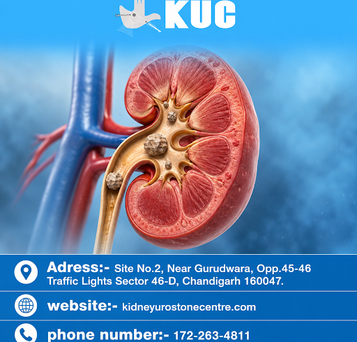
A kidney stone is a hard, crystalline mass that forms inside the kidney when certain dissolved minerals in urine — primarily calcium, oxalate, uric acid, or phosphate — become so concentrated that they crystallise rather than remain in solution. Stones range in size from a grain of sand to a golf ball. Most cause no symptoms until they begin to move.
When a stone moves from the kidney into the narrow tube connecting the kidney to the bladder — the ureter — it creates an obstruction. The kidney continues producing urine, which has nowhere to go. Pressure builds. The ureter contracts and spasms in an attempt to push the stone forward. The result is renal colic: a pain that comes in waves, intensifies without warning, and can radiate from the back and flank down to the groin and, in men, into the testes.
There is no comfortable position during renal colic. Unlike the pain of a pulled muscle, which eases with stillness, kidney stone pain typically does not respond to postural changes. It demands medical attention.
The Four Main Types of Kidney Stones
Understanding the type of stone a person forms matters. It determines the prevention strategy, the likelihood of recurrence, and in some cases the choice of treatment. According to StatPearls (NCBI), the distribution of stone types is as follows:
Calcium Oxalate and Calcium Phosphate Stones — 80 per cent of all cases
These are the most common kidney stones globally and in India. Calcium oxalate stones form when calcium and oxalate — a compound found in many plant foods — combine in concentrated urine. High dietary oxalate, low fluid intake, low dietary calcium (counterintuitively, restricting calcium increases stone risk by allowing more oxalate to reach the urine), and low urinary citrate all promote their formation.
Calcium phosphate stones form in more alkaline urine and are associated with conditions such as hyperparathyroidism and renal tubular acidosis.
Uric Acid Stones — 9 per cent of cases
Uric acid stones form when urine is persistently acidic. They are strongly associated with diets high in animal protein, gout, metabolic syndrome, obesity, and type 2 diabetes. Unlike calcium stones, uric acid stones are radiolucent — they do not show up on a plain X-ray — which means they can be missed on basic imaging and require ultrasound or CT for detection. They are more common in men than women and are increasingly prevalent in India as dietary patterns shift toward higher animal protein consumption.
Struvite Stones — 10 per cent of cases
Struvite stones, also called infection stones, form in alkaline urine as a direct consequence of chronic urinary tract infections caused by urease-producing bacteria, particularly Proteus mirabilis. These bacteria break down urea to produce ammonia, raising urine pH and creating the chemical conditions for struvite crystals to form. Struvite stones grow rapidly, can fill the entire kidney in a shape called a staghorn calculus, and are more common in women — who are already more prone to recurrent UTIs. They almost always require surgical intervention. Left untreated, they destroy kidney tissue and can cause life-threatening infection.
Cystine Stones — fewer than 1 per cent of cases
Cystine stones arise from a hereditary disorder called cystinuria, in which the kidneys fail to reabsorb the amino acid cystine properly, leading to its accumulation in urine. They tend to form early in life, recur frequently, and require lifelong management. Though rare, they are clinically significant because they are resistant to shock-wave lithotripsy and require specialist urological management.
India’s Stone Belt: A Geography of Risk
Kidney stone disease does not affect India uniformly. Published research in the journal Foods (PMC6352122) identified a clear geographic pattern: the states of Rajasthan, Gujarat, Maharashtra, Punjab, Haryana, and parts of Delhi constitute India’s “stone belt” — a zone of significantly elevated incidence driven by the convergence of climate, water quality, and dietary patterns.
In northern India, kidney stone prevalence reaches 15 per cent of the population. A study in the Indian Journal of Urology noted that stone belt states contribute to nearly one-third of all kidney stone cases reported nationally.
Three factors explain this pattern:
Hard groundwater: The Central Ground Water Board’s 2019 survey found calcium concentrations in parts of western Rajasthan exceeding 200 mg per litre — more than double the WHO’s recommended limit. High mineral content in drinking water directly increases the urinary load of stone-forming substances.
Climate: High ambient temperatures cause sweat-related fluid losses, concentrating urine and raising the mineral supersaturation that drives crystal formation. Climate projections published in PNAS suggest that warming temperatures will expand stone belt zones globally by 2050.
Diet: Stone belt populations consume higher quantities of animal protein relative to other Indian regions. Animal protein raises urinary uric acid and oxalate while reducing urinary citrate — three simultaneous changes that all promote stone formation. Research also notes low consumption of citrus fruits in these regions, removing a key dietary inhibitor of stone formation.
This is not a reason for fatalism. All three factors are either addressable or manageable with appropriate guidance.
Symptoms: What Kidney Stones Feel Like — and When They Are Silent
The majority of kidney stones cause no symptoms until they move. A stone resting in the kidney may sit undetected for months or years, discovered only incidentally on imaging performed for another reason.
When a stone begins to move, symptoms typically include:
Renal colic: Severe, wave-like pain in the flank, lower back, or side, below the ribs. Pain may radiate to the lower abdomen and groin. In men, it often extends into the testes or penis. In women, it can radiate to the vulva. The pain does not improve with changes in position.
Haematuria: Blood in the urine — visible as pink, red, or brownish discolouration — caused by the stone abrading the lining of the ureter or bladder. Blood may not always be visible to the naked eye; microscopic haematuria is detected on urine testing.
Urinary urgency and frequency: Stones near the bladder irritate the ureter-bladder junction, producing a persistent urge to urinate accompanied by small urine volumes.
Nausea and vomiting: The kidneys and digestive system share nerve pathways. Severe renal colic regularly triggers nausea and vomiting through visceral nerve connections, not because of gastrointestinal disease.
Fever and chills: These indicate infection. A stone causing obstruction with concurrent infection is a urological emergency. Infected, obstructed urine cannot drain, and bacteria multiply rapidly in a closed space. This can progress to sepsis within hours.
One important clinical note: men and women may interpret early symptoms differently. Women sometimes attribute early flank pain or urinary changes to menstrual cramps or a urinary tract infection, leading to delayed diagnosis. Men with uric acid stones may have existing gout and attribute early symptoms to their underlying condition. Both lead to the same outcome — a stone that has more time to grow, obstruct, and damage.
What Causes Kidney Stones to Form? The Biological Mechanism
Stone formation is not random. It occurs when urine becomes supersaturated with stone-forming substances — meaning more dissolved mineral is present than the urine can hold in solution. Several factors drive supersaturation:
Low fluid intake concentrates all urinary minerals, increasing the risk across all stone types.
Dietary excess: High animal protein raises urinary calcium, uric acid, and oxalate while reducing urinary citrate and pH. High sodium intake increases urinary calcium excretion. High fructose intake — increasingly common in processed foods — raises uric acid production and urinary oxalate.
Low dietary calcium: This is a common misconception. Restricting calcium in the diet does not reduce calcium stone risk. It increases it, because dietary calcium normally binds oxalate in the gut, preventing its absorption. Without adequate calcium intake, oxalate reaches the kidney in higher concentrations.
Genetics: A first-degree relative with kidney stones confers a 2.4 times higher risk of developing stones, according to research published in the Journal of Urology. Specific genetic disorders such as cystinuria and primary hyperoxaluria carry near-certain stone formation.
Medical conditions: Hyperparathyroidism, gout, type 2 diabetes, Crohn’s disease (which increases oxalate absorption), obesity, and recurrent UTIs all elevate kidney stone risk through distinct but documented mechanisms.
Medications: Some medications — including certain diuretics, calcium-based antacids, topiramate, and high-dose vitamin C supplementation — can promote stone formation as a side effect.
Prevention: What the Clinical Evidence Actually Shows
Kidney stone recurrence rates are high. Without intervention, between 27 and 50 per cent of patients experience a recurrence within five years, with annual recurrence rates of 15 to 20 per cent. Prevention is not optional for anyone who has formed a stone once.
Hydration — the most evidence-supported intervention
Drinking sufficient water to produce at least 2 to 2.5 litres of urine per day is the single most consistently supported preventive measure across all stone types. A Cochrane Database review and multiple clinical guidelines confirm that increasing water intake reduces recurrence. Urine should appear pale yellow throughout the day. Dark urine is a reliable warning sign of inadequate hydration. In India’s stone belt regions, particularly during summer months, 3 to 3.5 litres of daily fluid intake is appropriate.
Lemon juice and citrus fruits
This recommendation is not folk medicine — it has a strong evidence base. Citrate is a natural inhibitor of calcium stone formation. It binds calcium in the urine, reducing the amount available to crystallise with oxalate or phosphate. It also directly inhibits crystal growth and aggregation.
Lemon juice contains the highest concentration of citric acid among commonly consumed citrus fruits (49.2 g/kg). A randomised controlled trial published in eClinicalMedicine (The Lancet, 2021) demonstrated that 60 ml of fresh lemon juice twice daily, added to standard diet, reduced stone recurrence at one year with a hazard ratio of 0.43 — meaning stone recurrence was more than halved in the treatment group. Urinary citrate increased from an average of 142 mg/day to 346 mg/day in supplemented patients. A 2022–2023 randomised crossover trial (PMC10487295) confirmed that lemon-tomato juice supplementation significantly reduced established risk factors for calcium oxalate stone formation.
The practical application for Indian patients is simple: daily consumption of fresh lime or lemon juice, diluted in water, is a low-cost, evidence-supported protective measure with no significant adverse effects.
Dietary adjustments
Reduce animal protein to moderate quantities — not elimination, but reduction. Limit sodium intake, as sodium directly increases urinary calcium. Do not restrict dietary calcium; instead, consume calcium through food (dairy, fortified plant foods) with meals, so it can bind oxalate in the gut. Moderate oxalate-rich foods — spinach, nuts, tea, chocolate — particularly if prior stone analysis has confirmed calcium oxalate composition. Reduce fructose from processed foods and sugary drinks.
Pharmacological prevention
For patients with confirmed biochemical abnormalities, first-line pharmacological options include:
Potassium citrate for patients with documented low urinary citrate or acidic urine — it is more effective than lemon juice alone but carries gastrointestinal side effects that limit long-term adherence in approximately one-third of patients.
Thiazide diuretics to reduce urinary calcium excretion in patients with hypercalciuria.
Allopurinol to reduce uric acid production in patients with uric acid stones or hyperuricosuria.
These medications are recommended by both the American Urological Association (AUA) and the European Association of Urology (EAU) guidelines for patients with recurrent stones.
Treatment Options: From Waiting to Surgery
Treatment depends on stone size, location, composition (where known), and whether obstruction or infection is present.
Watchful waiting with medical expulsive therapy: Stones smaller than 5–6 mm have a reasonable probability of passing spontaneously. Alpha-blocker medications such as tamsulosin relax the ureteral smooth muscle, facilitating passage. Pain is managed with anti-inflammatories. This approach requires careful monitoring and is not appropriate when infection or significant obstruction is present.
Extracorporeal Shock Wave Lithotripsy (ESWL): High-energy shock waves applied from outside the body break the stone into fragments small enough to pass in urine. Most effective for stones up to 2 cm in the kidney or upper ureter. Not suitable for cystine stones, which absorb shock waves poorly, or for stones in the lower pole of the kidney, where fragment clearance is less reliable.
Ureteroscopy (URS): A thin flexible scope is passed through the urethra, bladder, and into the ureter or kidney. Stones are fragmented with laser energy and the pieces removed or allowed to pass. This is the most commonly used surgical approach, suitable for ureteral stones of most sizes and renal stones up to 2 cm.
Percutaneous Nephrolithotomy (PCNL): A small incision in the flank provides direct access to the kidney through the skin. A scope fragments and removes the stone under direct vision. This is the standard approach for large stones (greater than 2 cm), staghorn calculi, and stones in patients for whom ESWL or ureteroscopy have failed or are unsuitable.
Emergency intervention: A stone causing obstruction with concurrent infection is a urological emergency requiring immediate drainage — either by ureteral stent or percutaneous nephrostomy — to decompress the obstructed kidney before sepsis develops.
When to Seek Medical Care: A Practical Guide for Patients
| Symptom | Action |
|---|---|
| Mild back or flank pain, no fever | See a doctor within 24 hours |
| Blood in urine with or without pain | See a doctor promptly — do not wait |
| Severe pain — cannot find comfortable position | Go to emergency care |
| Fever, chills, with urinary symptoms | Emergency — this may be an infected stone |
| Nausea and vomiting with back pain | Emergency care |
| Unable to urinate despite urge | Emergency — possible complete obstruction |
| Previous stone history, new similar pain | Contact your urologist — do not assume it will pass |
Both men and women experience the same core symptoms. Men may have additional groin or testicular pain. Women sometimes attribute early symptoms to menstrual or gynaecological causes, which delays diagnosis. If in doubt, a simple urine test and ultrasound provide a rapid first-line assessment.
Recurrence: The Underestimated Threat
The most important thing to understand about kidney stones is that having one significantly increases the risk of having another. Without metabolic evaluation and targeted prevention, recurrence rates reach 50 per cent within five years. Each recurrence carries the risk of further renal damage, particularly when stones cause obstruction or are complicated by infection.
After passing or treating a stone, patients benefit significantly from:
A 24-hour urine collection to identify the specific metabolic abnormality driving stone formation — whether excess calcium, oxalate, uric acid, or low citrate.
Stone analysis, where fragments are collected and sent for chemical composition analysis. This directs prevention strategy more precisely than empiric treatment.
Follow-up imaging at defined intervals to detect silent stone growth before symptoms develop.
Conclusion: Information Is the First Line of Defence
Kidney stone disease is one of the most prevalent urological conditions in the world and in India. It is also one of the most preventable. The combination of adequate hydration, dietary adjustment, awareness of individual risk factors, and — where stones have already formed — specialist evaluation and targeted prevention can substantially reduce the burden this disease places on individuals, families, and the healthcare system.
The evidence is consistent across decades of research: behaviour matters more than genetics in most cases of kidney stone disease. This means the disease responds to what people know and what they choose to do with that knowledge.
Do not wait for the pain to make the decision for you.
This article is written for general public health awareness and does not constitute medical advice. Any person experiencing symptoms consistent with kidney stones should seek evaluation from a qualified urologist or nephrologist. For specialist kidney and urological stone care, visit kidneyurostonecentre.com